


Onychomicosis is a type of fungal lesion, which affects exclusively the surface of the nails.In order to prevent the spread of the infection over time, you should know how the nail fungus appears, since the therapeutic effect will be achieved faster if the treatment has started in an initial phase of infection.Very often, the disease occurs in the elderly because of the weak immunity, but the risk of obtaining a disease concerns each person.There is no single classification of damage to fungal nails;In medical practice, it is customary to distinguish them in the place of location and the depth they penetrate.The infection is also grouped according to the type of pathogen.
Types of pathogens and primary signs
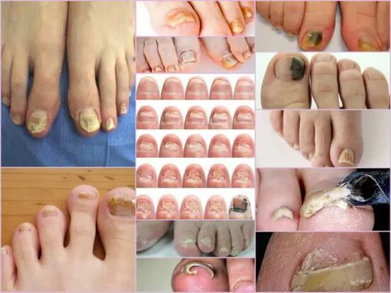
The symptoms of the nail fungus in the initial phase are easily determined directly from the nail conditions.This sign is the most instructive, since onychomycosis is always manifested in the form of changes in the color of the surface, deformation of the bed, exfoliation and any external changes.The latter are expressed by roughness, by the formation of grooves and cracks, as well as at interspersed points and general viorations of the nail.
The main sign of a healthy nail is pink and transparency.The onychomycosis in any stage is characterized by the turbidity of the nail and the change of color in yellow, brownish or black-greenish.In an advanced phase, the surface can acquire a black shade against the background of almost complete destruction.
The external signs of infection with a mushroom depend on the type of pathogen that has infected.In medical practice, the following possible lesions are distinguished:
- Candida infection with a mushroom, which is expressed by the unloading of the nail directly at the base of the box.The candidiasis of the rollers around the nail will be characteristic of candidiasis.This version of the complication can have a bacterial source in the form of streptococci or staphylococci, or expressed in the form of medium or psoriasis plates;
- Dermatophytes of the type of Trichophyton's rubbum.In this case, the penetration of the infection occurs directly from the free edge of the nail.The first symptoms of this pathogen are the appearance of a yellowish stain on the surface.In the field of neoplasm, the nails crumble and the point itself tends to increase.The commonplace of localization of the neoplasm is along the plate, parallel to the rollers for nails, in this case, the infection is called distal-lameral.There is another form of defeat by this pathogen - distal, in which the point appears on the part removed from the hole, mainly at the center of the free edge;
Important!
This type of mushroom occurs more often on the thumbs of the legs, gradually transforming the nails into a loose yellowish mass.In the absence of adequate treatment, the disease flows into hyperkeratosis.The nail plate is completely destroyed due to the spread of the infection around the perimeter.
- Dermatophytes like Trichophophyton Mentagophytes.The onychomycosis with these exciters is also based the most often based on the nails of the thumbs, less often than small fingers.An infection with a mushroom of this type requires a therapy not only of the nail, but also of the feet due to the rapid spread of the pathogen.A symptomatic disorder can be like Leikonichia, a common disease in medical practice.The main signs are the white spots that appear on various parts of the nail, neoplasms are distinguished by a non -standard and various size shape.It is easy to distinguish the mushroom from Leakonichia - in the latter case, the points are an air accumulation, which is not observed with fungal damage;
- Mushroom mushrooms.This damage option is significantly less often found than a candidate or dermatophytic form.The main sign of such an infection: the surface of the plate acquires a dark and almost black shadow.Not the entire finger can be completely infected, but only part of the nail plate.The first signs of nails mushrooms on the legs of this type are a strong change of color.The onychomycosis can develop in the form of a longitudinal strip of black or dark green in the background of the rest of the rose of the nail.
Diagnostics by type of change
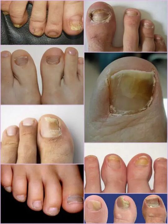
It is not difficult to note a nail fungus on the legs even in the primary phases of the infection, since an infection of this type occurs actively quite from the first day of the injury.Instead of the usual transparent nails of pale pink in the patient, a significant surface deformation and a general change in the condition are observed.The affected area has a matte opaque yellowish tint, which appears mainly on the thumbs.The type of mushroom and the degree of damage are the decisive factors.
In the first phase, the damage fungal in the legs on the legs is the appearance of small fires.The thickening of the dish and the keratinization of the bed under the nail will be characteristic.This phase is accompanied by a phenomenon as partial detachment and unloading of the nail plate, which acts from a source of infection for healthy people.
Despite the active thickening of the plate, its constant grounding can be observed, regardless of current factors.The characteristics of each stage and the symptoms of the mushroom on the legs depend directly on the type of pathogen.
Depending on the changes that occur with the nail plate, three options are distinguished for the onychomicosis:
- Hypertrophic;
- Normotrophic;
- Atrophic.
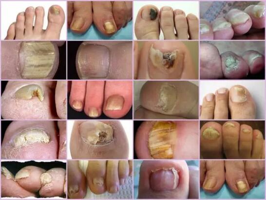
In the first case, there is a strong change in the shadow of the nail plate, its destruction along the edges and the deformation of the surface of the plate.The nail thickens so much that it causes discomfort and painful sensations during the walk.The normotrophical myrmo of the nails on the legs stands out for the presence of a healthy shine, but the plate itself acquires a little yellow, points are formed.With an atrophic type of damage, brown and gray outbreaks are formed on the surface of the nails.Thanks to them, it is possible to carefully determine the position of the pathogen.
Important!
The atrophic or on -niced type of nail mushrooms is characterized by thinning of the nail plate and not by its growth, as in other cases.Those areas on which the pathogen is located tend to detach themselves.The lack of adequate treatment leads to an advanced phase: the complete refusal of the nail plate.
Classification of location
Another sign with which it is possible to separate the fungal damage to the finger nails depends directly on the position of the outbreaks on the nail plate.This also includes the depth of the pathogen, which in turn allows the approximate duration of the next therapy and the possibilities of rapid recovery.
The fungal diseases of the legs of the legs in the location position are classified in the following groups:
- Onychomicosis is a type of white surface - the appearance of many white points on the surface of the nail plate.A fungal infection leads to the detachment of the skin in the places of appearance of points on which there is an active discharge of stairs.The advanced phase leads to the complete destruction and refusal of the plate;
- Distal - develops on the free nail edge.The color change is first observed on a small area of the plate, after which there is an active expansion of its borders.The lesion is characterized by a gray-yellow or brown color, as well as an irregular irregular surface and gradual exfoliation;
- The lateral onychomycosis has the phases of distal similar development, but is located exclusively on the lateral sides of the nail plate;
- Total infection - complete infection of the entire surface;
- Proximal onychomycosis.This disorder begins its activity from the cuticle, which is inflamed, so the mushroom quickly affects the nail and the process itself begins with the appearance of a small white spot near the inflamed area of the perolition roller.
The most common forms of mycosis of the nails on the legs are lateral and distal, which are usually caused by dermatophytes.These forms of damage such as proximal and white can act as a secondary disorder that accompanies the disease of the immune system, for example Vich.Total damage to the nail with a mushroom should be considered as an advanced phase of development of any mushroom under the nail.
Features of the mushroom with a microscope
Despite the presence of an impressive number of signs of onychomicosis, other disorders associated with skin problems are accepted and not of an infectious nature for fungal damage to the nails of the legs.It is possible to determine the exact diagnosis and the type of pathogen only in the laboratory under the microscope, for which, in a hospital, a scraping of biological material from the areas concerned is performed.
The resulting biomaterial is pre -piece in an alkaline environment, after which a multiple increase is performed.Studying the mushroom of the microscope nails allows you to see an active pathogen, the external shape of which determines its type, the distribution scale and an approximate degree of damage.On a nutritional medium, it is possible to predict the approximate growth of the colonies, which allows not only to make an accurate diagnosis, but also to determine the limitation of the infection.
Important!
Since it is possible to determine the presence of onychomicosis only in a laboratory, it is not necessary to postpone a visit to the doctor to the minimum suspicion.The nail fungus develops quickly and the waste of time increases therapy.
What is an alarming signal?
The fungus of the nails on the legs manifests itself from some symptoms that are partially similar to some skin disorders.The following signs are more likely to indicate an onychomomotic injury that requires medical intervention:
- The appearance of yellow spots on the plate, its deformation, a change in the structure, which has not been observed previously;
- The darkening of the dish, the loss of transparency, some photos of the nail fungus on the legs demonstrate a shadow close to black, which is characteristic of the molds of pathogens;
- Inspiration and keratinization of the nail plate or vice versa too sharp;
- Refusal of the nail from the bed, detachment of stairs and crumbs;
- Swelling of the roller, hanging it over the plate, release of liquid;
- The nails affected by the mushroom seem inflamed, regardless of the type of pathogen.
All these symptoms indicate a high risk of infection.Some external symptoms of modification of the nail plate structure may be the result of other diseases.With an increase in fragility, the quantity of calcium and iron in the body should be increased, an increased tuberosity means any body infection, with strips and purely white points, a lack of copper or zinc is possible.Despite the fact that in the photo the nail fungus resembles focal damage to the dish with a change of color closer to yellow, this does not always indicate the infection.Possible yellow can indicate various problems with the stomach and liver.
It is not difficult to recognize the onychomycosis on the fingers with external signs, despite the wide classification of the disease.But you can determine the exact type of pathogen and the damage stadium can only be in the laboratory.